

Outline
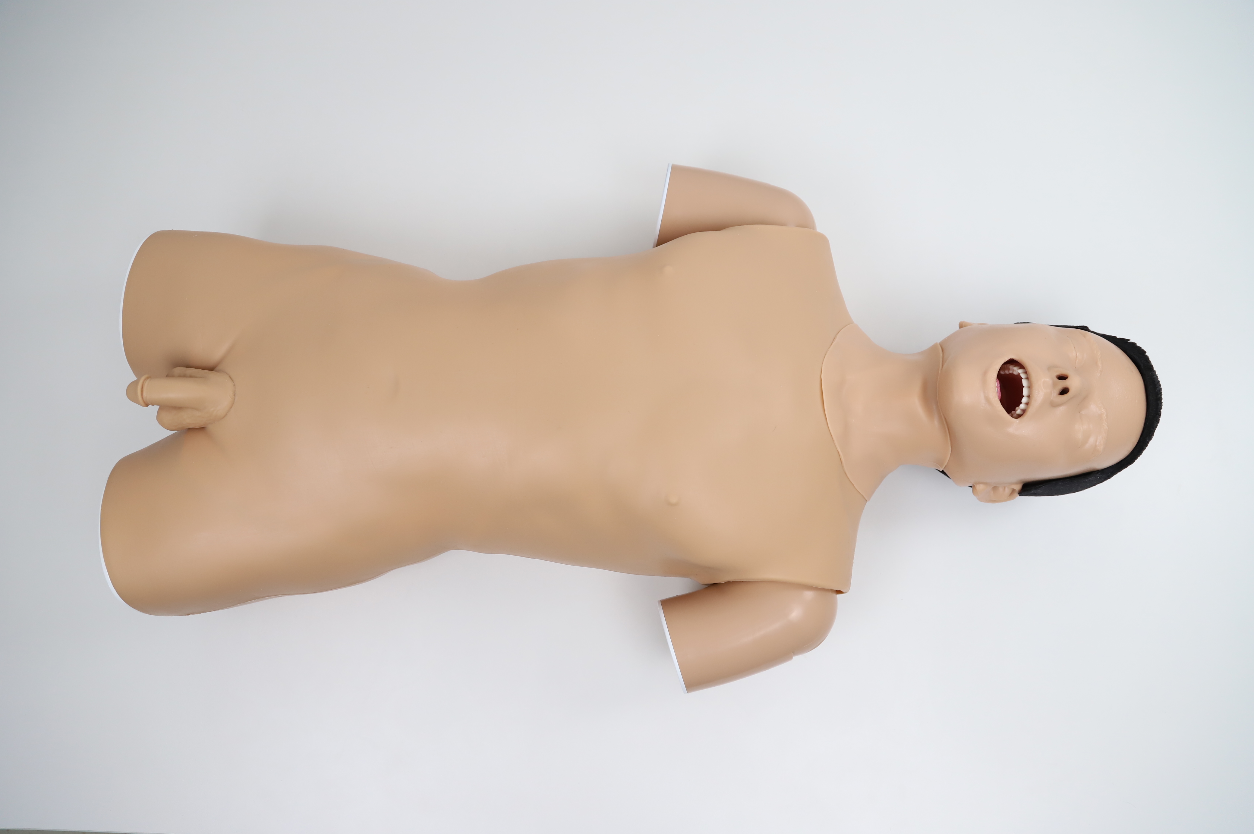
The Upper GI model features a male head, neck, and torso with anatomically accurate representations, providing a highly realistic simulation of upper GI endoscopy training.
Skills Gained
· Identification of upper GI anatomical structures
· Fiberoptic endoscopy techniques
· Endoscopic Retrograde Cholangiopancreatography (ERCP)
· Endoscopy techniques
Features
· Anatomy:
1) Nasal cavity, oral cavity, pharynx, tonsil, palatoglossal arch, palatopharyngeal arch, pyriform recess, epiglottis, tracheal opening, esophageal opening, esophagus, stomach and duodenum
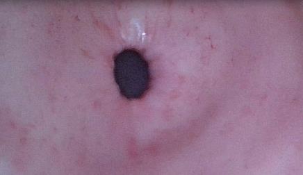
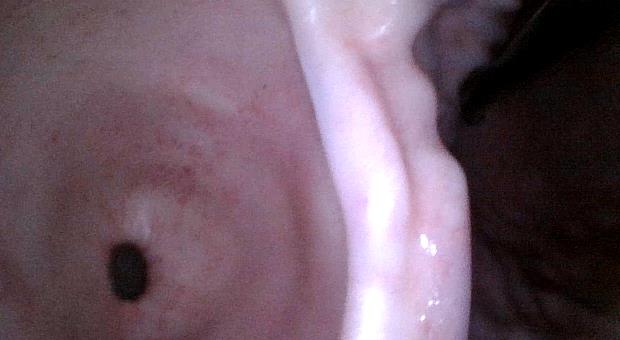
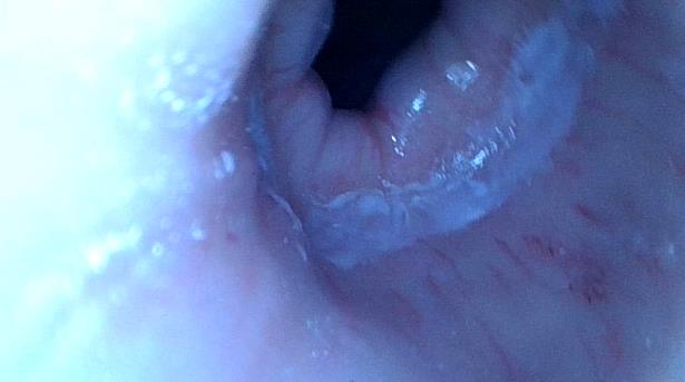
2) Realistic and complete internal structures including cardia, dentate line, pylorus, major duodenal papilla (papilla of Vater), accessory duodenal papilla, and gastric angle. The main papilla contains part of the bile duct and pancreatic duct
· Key Features:
1) The simulated upper GI structure derives from CT scan of a real person
2) The model’s soft material allows clear endoscopic view of capillary networks with realistic color, texture, and tactile feedback
3) The simulated stomach allows for deflation and inflation maneuvers
Previous: Lower GI Endoscopy Training Model
Stock code :833047
Address:2nd & 3rd Floor, West 6th Building, 18 West HaiTai Road, Tianjin, China
Postcode:300384
Phone:4006-355-510
+86-22-83711066
Fax:+86-22-83711065
Email:info@tellyes.com

 Endoscopic